In September 2008 the Canterbury Dental Service (CDS) introduced digital radiography for its patients. This replaced conventional radiographs (a wet film taken at the clinic which is then sent to a dark room for processing) which had been used since the 1920s.
The new digital image can be entered into a new electronic oral health record (Titanium) which was also introduced in 2008.
CDS provides oral care for approximately 77,000 Canterbury children. The new digital imaging system is now very much a part of the daily oral health care for these children. Therefore it is essential to ensure that the diagnostic x-ray facilities are providing consistent, high quality images with a minimum of exposure to patients and staff. This is the clinical motivation for quality control checks on equipment, but in New Zealand this is also backed-up with regulatory requirements for all human radiographic imaging to be part of a regular quality assurance programme.
CDHB Clinical Director, School & Community Dental Service Martin Lee and Public Health Dentist Tule Fanakava Misa approached the Medical Physics Bioengineering (MPBE) team for their help in developing systems to test the three components involved in the new digital imaging system. These components are the x-ray generator, plate and scanner, and computer/monitor on which the images are displayed.
Tule teamed up with Nick Cook, MPBE Imaging Scientist to find a solution. He developed a quality assurance programme which monitors performance and function of these components on a regular basis. Central to this new quality assurance programme was a special device, the Quality Assurance Phantom (QA Phantom) that could simulate the demanding aspects of dental x-ray imaging and test the true performance of equipment for clinical imaging. Although the final phantom was simple in design and low cost compared to commercial products, it required precision machining in the MPBE Bioengineering workshop to achieve the required performance.
The phantom consists of a small block of Perspex (PMMA) with a series of different size and depth holes. The Perspex box is placed over an aluminium plate to mimic a tooth with subtle density changes. MPBE also constructed a special protective carry case for the phantoms.
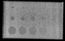
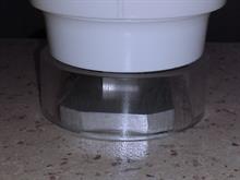 The QA Phantom is located inside a Perspex cylinder that achieves reproducible positioning for repeated x-ray measurements. The x-ray image of the phantom is checked on a calibrated monitor to see how many of the holes are visible and this allows close monitoring of the performance of the machines to correct any problems before they affect patient images.
 Each dental clinic and mobile dental facility now has a QA Phantom device so that trained staff can perform monthly checks of the equipment. These checks ensure that the digital dental images are accurate and achieve the performance require to catch any dental problems before they lead to bigger issues. They have been used since beginning of the third school term 2014. Staff have been trained to use the device and do their own checks monthly.
The QA Phantom is used in Christchurch Hospital and community dental clinics but has not been adopted elsewhere in New Zealand as yet because not all dental clinics have switched to digital images.